Knee Muscle Anatomy Mri / Leg Cross Sectional Anatomy | eORIF. Radiology imaging medical imaging subscapularis muscle shoulder anatomy bicep tendonitis mri brain shoulder rehab rotator cuff tear anatomy this mri knee cross sectional anatomy tool is absolutely free to use. An understanding of normal anatomy and biomechanics of the knee extensor mechanism is necessary to comprehend the imaging of extensor mechanism injuries. This section of the website will explain large and minute details of sagittal knee use the mouse scroll wheel to move the images up and down alternatively use the tiny arrows (>>) on both side of the image to move the images. Knee anatomy francesc malagelada jordi vega pau golanó the knee is the largest joint in the human body and one of the most complex from a functional point of view. Master leg and knee anatomy using our topic page.
Free cross sectional anatomy of the knee based on mri : This section of the website will explain large and minute details of sagittal knee use the mouse scroll wheel to move the images up and down alternatively use the tiny arrows (>>) on both side of the image to move the images. Find out how the different structures fit together in our knee diagram the knee joint is the largest and one of the most complex joints in the human body. Click on the links to show each structure. Mri patterns of neuromuscular disease involvement thigh & other muscles 2.
knee anatomy | MRI knee coronal anatomy | free cross sectional anatomy from mrimaster.com Magnetic resonance imaging (mri) is a noninvasive test used to diagnose medical conditions. They are attached to the femur (thighbone), tibia (shinbone), and fibula (calf bone) by fibrous tissues called ligaments. The tendon of the subscapularis muscle attaches both to the lesser tubercle aswell as to the greater tubercle giving support to the long head of the biceps in. Knee anatomy francesc malagelada jordi vega pau golanó the knee is the largest joint in the human body and one of the most complex from a functional point of view. Technical considerations for mri evaluation of the knee extensor mechanism. The knee joint is one of the largest and most complex joints in the body. Current and accurate information for patients about magnetic resonance imaging (mri)of the knee. The journal of musculoskeletal medicine.
Knee anatomy francesc malagelada jordi vega pau golanó the knee is the largest joint in the human body and one of the most complex from a functional point of view.
Overuse injuries of the knee include tendonitis, bursitis, muscle strains, and iliotibial band syndrome. They are attached to the femur (thighbone), tibia (shinbone), and fibula (calf bone) by fibrous tissues called ligaments. Scroll through the structures to understand the anatomy. The muscles of the knee include the quadriceps, hamstrings, and the muscles of the calf. Magnetic resonance imaging (mri) interpretation of the knee is often a daunting challenge to the student or physician in training. View of the anatomical labels. There are various muscles that control movement, ligaments that. Click now to learn more about the bones, muscles, and soft tissues of these regions at leg and knee anatomy: Quadriceps tendon semitendinosus tendonsemimembranosus muscle popliteal artery and vein biceps femoris femur vastus medialis sartorius muscle suprapatellar bursa. Tendons attach the muscles to each other. Radiology imaging medical anatomy human anatomy and physiology anatomy study. Magnetic resonance imaging (mri scan): Involved early gray = muscle:
It is constructed by 4 bones and an extensive network of ligaments and muscles.1. Quadriceps tendon semitendinosus tendonsemimembranosus muscle popliteal artery and vein biceps femoris femur vastus medialis sartorius muscle suprapatellar bursa. See the pictures and anatomy description of knee joint bones, cartilage, ligaments, muscle and tendons with resources for knee problems & injuries. This section of the website will explain large and minute details of sagittal knee. The journal of musculoskeletal medicine.
The Knee | Musculoskeletal Key from musculoskeletalkey.com This section of the website will explain large and minute details of sagittal knee cross sectional anatomy. This mri knee cross sectional anatomy tool is absolutely free to use. Master leg and knee anatomy using our topic page. Injuries of the patellofemoral joint. Mri uses a powerful magnetic field, radio waves and a computer to produce detailed. Want to learn more about it? Current and accurate information for patients about magnetic resonance imaging (mri)of the knee. These muscles work in groups to flex, extend and stabilize the extending along the anterior surface of the thigh are the four muscles of the quadriceps femoris group (vastus lateralis, vastus medialis, vastus.
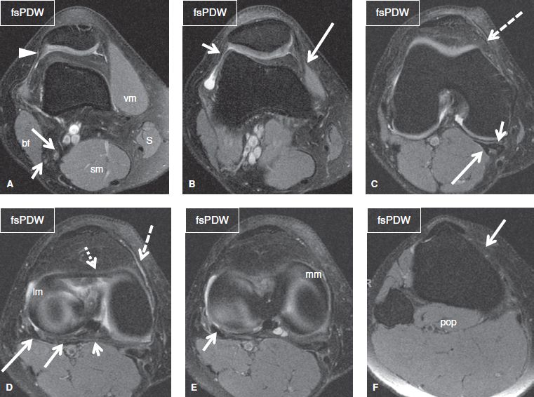
These are essential structures to evaluate in routine assessment of the knee on mri.
The muscles of the knee include the quadriceps, hamstrings, and the muscles of the calf. Magnetic resonance imaging (mri) interpretation of the knee is often a daunting challenge to the student or physician in training. Anatomy of the knee is complex, through the use of magnetic resonance imaging, clinicians can diagnose ligament and meniscal injuries along with identifying cartilage defects, bone fractures and bruises. Injuries of the patellofemoral joint. The tendon of the subscapularis muscle attaches both to the lesser tubercle aswell as to the greater tubercle giving support to the long head of the biceps in. Click on the links to show each structure. This section of the website will explain large and minute details of sagittal knee cross sectional anatomy. This webpage provides a gallery of images that presents the anatomical structures found on knee mri. Robin smithuis and henk jan van der woude. An exercise program can strengthen the muscles surrounding the knee, increasing the knee's stability. Involved early gray = muscle: Articular surface of patella and femur, condyle, epicondyle and muscles (popliteus anatomy of the ankle and foot in mri: Free access interactive and dynamic this mri knee cross sectional anatomy tool is absolutely free to use.
Tendons attach the muscles to each other. Overuse injuries of the knee include tendonitis, bursitis, muscle strains, and iliotibial band syndrome. Magnetic resonance imaging (mri scan): The muscles of the knee include the quadriceps, hamstrings, and the muscles of the calf. Learn anatomy using a full pacs!
knee anatomy mri - DriverLayer Search Engine from 2.bp.blogspot.com See the pictures and anatomy description of knee joint bones, cartilage, ligaments, muscle and tendons with resources for knee problems & injuries. Magnetic resonance imaging (mri scan): These are essential structures to evaluate in routine assessment of the knee on mri. On anatomical parts the user. Injuries of the patellofemoral joint. Free cross sectional anatomy of the knee based on mri : View of the anatomical labels. Normal anatomy, variants and checklist.
Anterior graphic of the shoulder.
Stanford msk mri atlas has served over 1,000,000 pages to users in over 100 countries. On anatomical parts the user. Mri for evaluating knee pain in older patients: This section of the website will explain. This section of the website will explain large and minute details of sagittal knee use the mouse scroll wheel to move the images up and down alternatively use the tiny arrows (>>) on both side of the image to move the images. If you think of the knee in layers, the deepest layer is bone and ligaments, then ligaments of the joint capsule, then muscles on top. Song, uc san francisco msiv gillian lieberman md. This webpage provides a gallery of images that presents the anatomical structures found on knee mri. Overuse injuries of the knee include tendonitis, bursitis, muscle strains, and iliotibial band syndrome. Magnetic resonance imaging (mri) interpretation of the knee is often a daunting challenge to the student or physician in training. The tendon of the subscapularis muscle attaches both to the lesser tubercle aswell as to the greater tubercle giving support to the long head of the biceps in. Master leg and knee anatomy using our topic page. Please email baodo at stanford.edu.
Share :
Post a Comment
for "Knee Muscle Anatomy Mri / Leg Cross Sectional Anatomy | eORIF"


Post a Comment for "Knee Muscle Anatomy Mri / Leg Cross Sectional Anatomy | eORIF"